Mariana Grecu, Oana Tănase, Mădălina Elena Henea, Valentin Nastasă, Cristina Mihaela Rîmbu
ABSTRACT. This study assessed seasonal incidence, economic losses, the efficacy of therapeutic protocols, the recovery time of affected animals and specific prophylactic measures applied to sheep with acute infectious pododermatitis. The studies were conducted over a period of 12 months in 3 different sheep farms from private units in the same area. The results of the study showed an increased incidence of the disease in all 3 farms, with an average of 26.94% of the sheep flock. The incidence of the disease was increased in the months of April-May-June-July and September-October (30%), when there were heavy rains. The high morbidity led to economic losses through the decrease in milk production by approximately 30% and the decrease in the weight of the sheep by 10.58% (4.2 kg) of their normal weight. The therapeutic protocol applied locally as well as parenterally, combined with a foot bath with 10% zinc sulphate solution, were effective in treating acute infectious pododermatitis of sheep. The average recovery time (days) was approximately the same in the three groups of sheep (5.25 ± 0.68 days for cases with moderate diseases and 10.2 ± 0.22 for cases with severe diseases).
Keywords: acute infectious pododermatitis; sheep; Fusobacterium necrophorum; Dichelobacter nodosus.
Cite
ALSE and ACS Style
Grecu, M.; Tănase, O.; Henea, M.E.; Nastasă, V.; Rîmbu, C.M. Prevalence, economic impact and therapeutic efficacy in acute infectious pododermatitis in sheep. Journal of Applied Life Sciences and Environment 2022, 55 (2), 233-243.
https://doi.org/10.46909/alse-552060
AMA Style
Grecu M, Tănase O, Henea ME, Nastasă V, Rîmbu CM. Prevalence, economic impact and therapeutic efficacy in acute infectious pododermatitis in sheep. Journal of Applied Life Sciences and Environment. 2022; 55 (2): 233-243.
https://doi.org/10.46909/alse-552060
Chicago/Turabian Style
Grecu, Mariana, Oana Tănase, Mădălina Elena Henea, Valentin Nastasă, and Cristina Mihaela Rîmbu. 2022. “Prevalence, economic impact and therapeutic efficacy in acute infectious pododermatitis in sheep” Journal of Applied Life Sciences and Environment 55, no. 2: 233-243.
https://doi.org/10.46909/alse-552060
View full article (HTML)
Prevalence, Economic Impact and Therapeutic Efficacy in Acute Infectious Pododermatitis in Sheep
Mariana GRECU*, Oana TĂNASE, Mădălina Elena HENEA, Valentin NASTASĂ and Cristina Mihaela RÎMBU
Iasi University of Life Sciences, Faculty of Veterinary Medicine, Department Preclinics, 8, Mihail Sadoveanu Alley, 700489, Iasi, Romania; e-mail: otanase@uaiasi.ro; madalina.henea@yahoo.com; vnastasa@uaiasi.ro; crimbu@uaiasi.ro
*Correspondence: mgrecu@uaiasi.ro
Received: Dec. 08, 2022. Revised: Dec. 29, 2022. Accepted: Jan. 02, 2023. Published online: Feb. 09, 2023
ABSTRACT. This study assessed seasonal incidence, economic losses, the efficacy of therapeutic protocols, the recovery time of affected animals and specific prophylactic measures applied to sheep with acute infectious pododermatitis. The studies were conducted over a period of 12 months in 3 different sheep farms from private units in the same area. The results of the study showed an increased incidence of the disease in all 3 farms, with an average of 26.94% of the sheep flock. The incidence of the disease was increased in the months of April-May-June-July and September-October (30%), when there were heavy rains. The high morbidity led to economic losses through the decrease in milk production by approximately 30% and the decrease in the weight of the sheep by 10.58% (4.2 kg) of their normal weight. The therapeutic protocol applied locally as well as parenterally, combined with a foot bath with 10% zinc sulphate solution, were effective in treating acute infectious pododermatitis of sheep. The average recovery time (days) was approximately the same in the three groups of sheep (5.25 ± 0.68 days for cases with moderate diseases and 10.2 ± 0.22 for cases with severe diseases).
Keywords: acute infectious pododermatitis; sheep; Fusobacterium necrophorum; Dichelobacter nodosus.
INTRODUCTION
Sheep breeding in Romania is a very old and important branch, with a tradition of centuries. Sheep represent the species that best capitalises on pastures and gives diversified productions with special biological value (Fesseha, 2021).
Infectious pododermatitis of sheep is a disease that causes significant economic losses to sheep producers. Infectious pododermatitis is a common condition in sheep and is an infectious and contagious disease that can cause great damage in sheep flocks around the world (Angell et al., 2015; Ansari et al., 2014; Raadsma and Dhungyel, 2013).
Infectious pododermatitis in sheep causes major economic losses on farms by reducing milk production (Duncan et al., 2014), wool yield and weight loss (Marshall et al., 1991; Nieuwhof et al., 2008; Rader et al., 2011). In addition, untreated sheep infect other animals in the flock, resulting in high mortality. Purulent necrosis, infections and superinfections that occur in the affected area lead to severe damage to the limbs, so that the animal stops moving and stops consuming food and water. Because of superinfections, septicaemia can occur, as can death of the animal (Angell et al., 2015; Raadsma and Dhungyel, 2013).
The disease is not usually fatal, provided that treatment is given in the early stages of disease, but deaths may occur in lambs of better breeds (Zanolari et al., 2021). Morbidity is thought to vary from 2–3% to 40–50%, which is related to predisposing factors (Davies et al., 1999). The more severe the course of the disease, the higher the morbidity can be, sometimes even reaching 100% (Swarnkar et al., 2021).
The disease is caused by an anaerobic germ, Dichelobacter nodosus, formerly known as Fusiformis nodosus, alone or in association with other anaerobic germs, of which Fusobacterium necrophorum is the most commonly involved (Belloy et al., 2007; Moore et al., 2005; Wilson-Welder et al., 2015), although these two anaerobic bacteria act synergistically and both are always involved (Clifton et al., 2019).
The incubation period varies with the season but is generally two weeks. Symptomatology is highly variable and depends on several factors such as soil conditions, the virulence of bacterial strains, the number of limbs affected, the duration of illness and secondary complications (Gelasakis et al., 2017).
Infectious pododermatitis of sheep is manifested by pain in the hoof, soft and warm hooves in the area where the infection is located, difficulties with movement and pain followed by lameness (Davies et al., 1999). In severe cases, sheep avoid movement and remain in a decubitus position without the opportunity to feed or water.
Sheep of all ages and breeds are susceptible to the disease, but adult females and rams are most susceptible, especially advanced breeds in which the disease is highly progressive (Gelasakis et al., 2017; Raadsma and Dhungyel, 2013). Rustic breeds and young sheep are more resistant because the disease has a benign course (Raadsma and Dhungyel, 2013). Factors favouring the disease include grazing on wet or marshy ground, sick and pregnant sheep, as the disease is highly contagious, prolonged grazing on wet bedding, a lack of foot hygiene and a lack of prophylaxis. Meteorological factors can cause the disease to appear and maintain in sheep farms. Thus, heavy rains during the year can influence the contamination of the environment in which the sheep live (Abbott and Egerton, 2003; Duncan et al., 2014). Predisposing factors include race, age, sex, growth system, excessive moisture, rich vegetation, heavy rainfall, ambient temperature, degree of damage to the interdigital integument and transhumance (Ansari et al., 2014). The disease takes a progressive course and, if untreated, leads to necrosis with the destruction of both the interdigital integument above the vertex and the sole integument. Purulent necrosis, infection and superinfection spreading to the area can also lead to exongulation, septicaemia and even death (Gelasakis et al., 2017).
Pododermatitis is an endemic disease in sheep flocks all over the world, including in Romania. Since sheep are raised in private farm-type units in Romania, the epidemiological situation of infectious pododermatitis is not concretely known.
Disease eradication management is impossible to apply because the D. nodosus strain was detected in the absence of lesions in sheep and other species (Ardüser et al., 2020), making this a factor favouring the permanent occurrence of the disease. As measures to reduce the incidence, permanent supervision of sheep flocks through periodic clinical examinations and the application of specific prophylaxis measures are considered.
The main aim of the study was to estimate the prevalence of acute infectious pododermatitis in sheep and the economic impact of this disease in sheep flocks in the area of Romania, where farmers are constantly faced with this condition.
MATERIALS AND METHODS
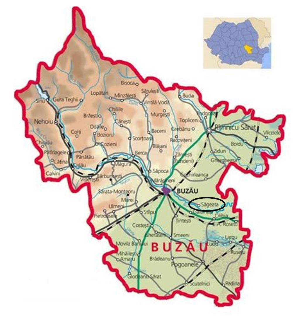
The studies were conducted between September 2021 and September 2022 on a total of 1721 sheep of Turcana breed of different ages, both adult sheep and youth (lambs), from 3 flocks in 3 different localities in Buzau County (Figure 1), which were constantly confronted with limb diseases in sheep. The flocks were from individual farms with more than 20 years of experience in sheep breeding and with more than 500 animals in each of the flocks, which were divided into 3 groups according to locality. For ethical reasons, we have named them: S1 with a flock size of 590 sheep, S2 with a flock size of 553 sheep and S3 with a flock size of 578 sheep. The sheep were raised in a traditional system, grazing during the day and stabled at night, with grazing in the summer and stabling in the winter.
The entire flock of sheep on each farm was examined every month during the study period to assess the seasonal incidence of the disease. Sick animals were isolated from healthy animals and kept on clean, dry and constantly changed bedding. Samples were taken from the affected limbs of sheep with severe lesions for bacteriological examination.
The local treatment was identical for all sheep with infectious pododermatitis from the 3 flocks, but the injection treatment was different, selecting 3 antibiotics for use in infectious pododermatitis in sheep: Gamithromycin 6 mg/kg for group S1, Tulathromycin 2.5 mg/kg for group S2 and Tilmicosin 10 mg/kg for group S3. The local treatment for all affected sheep was performed by mechanical cleaning, adjusting the nails with the removal of necrotic tissue if necessary, washing the lesions with 3% hydrogen peroxide solution and the local application of oxytetracycline spray (Terramycin® – Pfizer) for 7 days. Foot baths containing zinc sulphate 10% solution were used daily for at least 5 minutes for each sick sheep.
Thus, gamithromycin (commercial product Zactran® 150 mg/ml, Merial) was administered subcutaneously to sheep from farm S1, tulathromycin (commercial product Tuloxxin® 100 mg/ml, KRKA) was administered intramuscularly to sheep from farm S2 and tilmicosin (Tilmisone® 300 mg/ml, Mevet) was used subcutaneously in sheep from farm S3 (Table 1). The injectable antibiotics were selected based on their specifications for use in infectious pododermatitis in sheep associated with the virulent Dichelobacter nodosus and Fusobacterium necrophorum requiring systemic treatment as either single use or for a maximum of 2 days to avoid repeated administration and the possible emergence of antibiotic resistance, considering that the owners face this disease annually.
RESULTS AND DISCUSSION
Clinical manifestations of acute infectious pododermatitis were only observed in adult sheep, both females and males, with females predominating because their proportion in the flock was much higher.
Of the total 1721 sheep, acute infectious pododermatitis was diagnosed in about 30%, although preventive measures with 5% copper sulphate solution and 2% formaldehyde were applied.
Clinically manifested symptoms were movement difficulties, lameness causing lagging behind the herd, refusal to move in severe cases, pain and sensitivity of the affected area, apathy, loss of appetite, decreased milk secretion and obvious emaciation.
Table 1
Design for the therapeutic regimen of acute foot rot in sheep
|
Group |
Total number of animals/ herd |
Therapeutic protocol for sheep with acute infectious pododermatitis |
||||
|
Local treatment |
Duration of treatment (Day) |
Systemic treatment |
Dose and route of administration |
Duration of treatment (Day) |
||
|
S1 |
590 |
Hydrogen peroxide 3% + Oxytetracycline spray (Terramycin® – Pfizer) + Zinc sulphate 10% |
7 |
Gamithromycin (Zactran® 150 mg/ml, Merial) |
6.0 mg /kg, subcutaneous (1ml/25 kg) |
2 |
|
S2 |
553 |
Tulathromycin (Tuloxxin® 100mg/ml, KRKA) |
2.5 mg/kg intramuscular (1 ml/40 kg) |
2 |
||
|
S3 |
578 |
Tilmicosin (Tilmisone® 300mg/ml, Mevet) |
10 mg/kg subcutaneous (1ml/30 kg) |
2 |
||
Examination of the affected limb(s) revealed congestion and superficial necrosis of the interdigital integument with foul-smelling exudate, the erosion of adjacent tissue and erosion of the keratogenic membrane. Necrosis of the skin at the junction of the hoof capsule and in some cases the partial extension of the lesion with damage to the skin above the hoof was also found. Bacteriological examination revealed Dichelobacter nodosus and Fusobacterium necrophorum, bacteria which are always found in sheep with infectious pododermatitis.
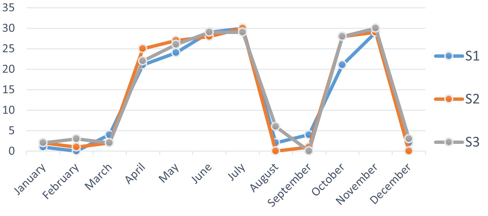
From the observations made, the incidence of pododermatitis in sheep was shown to record an average of 26.94% of the total flock of the 3 farms. The maximum morbidity was observed between April and June and between October and November (29–30%), periods during which rainfall was abundant. There were no cases of pododermatitis (Figure 2) in January, February and December when sheep were stabled and in August-September when precipitation was low in quantity (1–3%).
These results clearly show that one of the factors that favour the occurrence of the disease are wet soils and the high contagiousness of the disease. The high incidence of pododermatitis in sheep in the above months can be explained by the fact that soil moisture, due to abundant and prolonged rainfall in spring-summer and autumn, is highly conducive to maceration of the skin and horn of the nails, creating optimal conditions for the penetration and active multiplication of pathogenic microbial flora in the acropodial tissue.
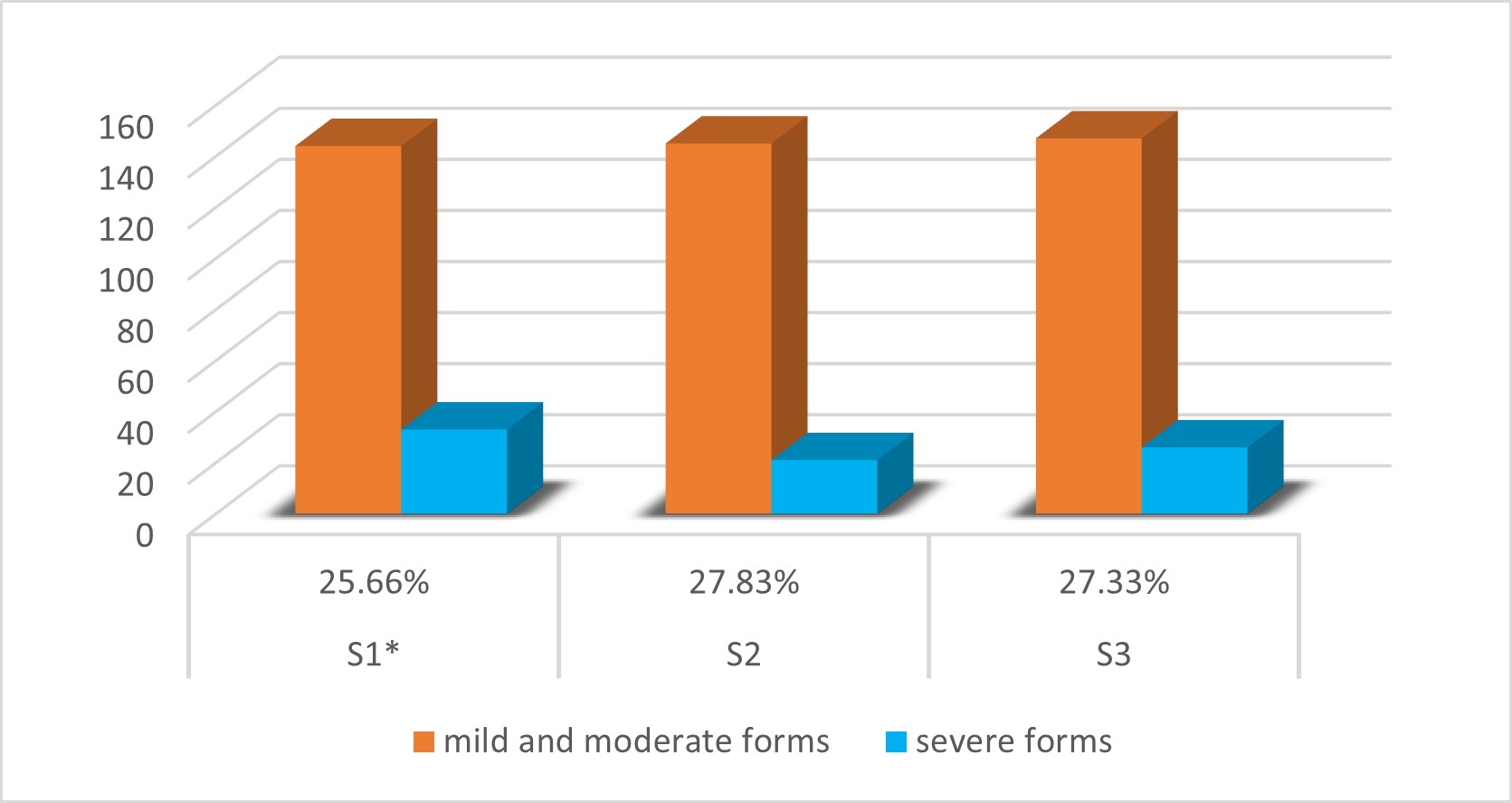
From the 12-month investigations (September 2021–September 2022), it appeared that about 30% of the total 1721 sheep developed the disease during one of the periods when it rained heavily and frequently. Thus, 177 sheep were recorded in the S1 pen (144 with mild or moderate forms and 33 with severe forms), 166 sheep in the S2 pen (145 with mild or moderate forms and 21 with severe forms) and 173 sheep in the S3 pen (147 with mild or moderate forms and 26 with severe forms) (Figure 3).
Of the total affected sheep (n=516), 53.5% (276/516) had a lesion on one limb, 37.26% (192/516) had two lesions on two limbs, 7.32% (38/516) had lesions on three limbs and only 1.92% (10/516) had lesions on all four limbs. The majority of sheep in the three flocks showed the hind limbs to be most severely affected.
When estimating the economic losses suffered by farmers, it was found that they were directly proportional to the percentage of diseased sheep and resulted in a significant decrease in milk production (Table 2), despite being in peak lactation.
Animals with acropodial lesions showed a decrease in milk production of about 30% on the days of peak disease development, depending on the severity of the lesions, although they were kept under the same feeding conditions as healthy ewes, which can be explained by painful suffering with the impossibility of movement, listlessness and inappetence.
Mean±SE values of weight in kilograms for all sheep were 39.5±0.5.
Table 2
Average decrease in milk production in sheep with infectious pododermatitis
|
Specification |
Average normal production/ sheep/day |
Sheep with minor or medium lesions |
Sheep with major lesions |
|
|||||||
|
|
S1 |
S2 |
S3 |
S1 |
S2 |
S3 |
S1 |
S2 |
S3 |
||
|
Mean daily milk production (ml) |
880 ± 0.10 |
890 ± 0.10 |
880 ± 0.10 |
– |
– |
– |
– |
– |
– |
||
|
Decrease in milk production/day |
ml |
– |
– |
– |
160 ± 0.10 |
180 ± 0.10 |
180 ± 0.10 |
260 ± 0.10 |
250 ± 0.10 |
250 ± 0.10 |
|
|
% |
– |
– |
– |
18.18 |
20.22 |
20.45 |
29.54 |
28.08 |
28.41 |
||
When the recorded data were analysed, greater emaciation was observed in animals with larger lesions than in animals with smaller lesions. Thus, a maximum weight loss of 4.2 kg (10.58%) and a minimum of 1.9 kg (4.81%) was observed in sheep with larger lesions of acute infectious pododermatitis compared with healthy sheep (Table 3).
The sick sheep were individually weighed together with the clinical examination to confirm the diagnosis. An average weight in kg was calculated between the healthy sheep and the sick sheep, after which the healthy sheep were weighed as a proportion of 30% of the herd from each farm.
This debilitation can be explained by increased pain sensitivity leading to the inability to move and eat. Local treatment with antibiotic and antiseptic sprays, daily immersion of the limbs in a 10% zinc sulphate solution for a minimum of 5 minutes and the subcutaneous or intramuscular injection of a broad-spectrum antibiotic have demonstrated efficacy and the necessity of therapy for this condition.
The injectable antibiotics gamithromycin, tulathromycin and tilmicosin were selected for therapy for several reasons: they are relatively new to therapy; they require a single administration, eliminating the stress of therapy and recommending a 24-hour repeat only for larger lesions; they are formulated specifically for this disease in sheep; and their antibiotic resistance is not yet known, considering that farmers regularly encounter this disease.
Administration of the therapeutic agents used in the 3 flocks of sheep resulted in a gradual improvement of the condition, such that a favourable response to treatment was observed in sheep with mild lesions and those with moderate lesions from the third day onward. Recovery was more than 95% of the treated cases after 5 days of drug application, but complete recovery occurred only after 10 days. In sheep with severe forms of the disease, an improvement in lesions was observed in more than 90% of the treated animals after 9–10 days of therapy and complete recovery occurred after 17–20 days. Mean recovery time (days) was approximately the same in all three flocks, both sheep with minor and moderate injuries (5.25 ± 0.38) and sheep with major injuries (10.2 ± 0.22). Sheep with minor or moderate injuries responded much more quickly than sheep with major injuries because they only had lesions on one or rarely two limbs.
A withdrawal period for milk and slaughter of 16–26 days was required in both lactating and meat sheep. Once the sheep had fully recovered, they were reintroduced into the flock with regular prophylactic use of disinfectant solutions containing 5% copper sulphate and 10% zinc sulphate.
Each of the injectable antibiotics used had a good therapeutic effect with a prolonged duration of action without causing adverse reactions in the sheep. The effectiveness of the injectable antibiotics used in our study is also reported by Strobel et al. (2014), via a comparative study of gamithromycin and oxytetracycline used in limb diseases in sheep, where they observed that gamithromycin was significantly more effective than oxytetracycline. In the case of tulathromycin, Mladenov et al. (2020), through the information provided, demonstrated the high sensitivity of the tested pathogens to tulathromycin and the high clinical and economic effect that is effective with a single dose of 2.5 mg/kg administered to small ruminants. Angell et al. (2015), in their study to evaluate the therapeutic effect of tilmicosin, demonstrated the reduction in prevalence and the clinical elimination of sheep with contagious digital dermatitis from the flocks studied in the UK.
The results of the bacteriological examination in our study, by isolating strains of Dichelobacter nodosus and Fusobacterium necrophorum from sick sheep, were similar to the results of various research studies that isolated these strains at a percentage of over 50% in the examined sheep (Ardüser et al., 2020; Belloy et al., 2007; Clifton et al., 2019; Fesseha, 2021).
Every farmer is aware of the fact that he must ensure the health status of the animals on his own farm, with the aim of ensuring the conditions of well-being and reducing the losses caused by diseases specific to sheep. However, this is impossible to achieve most of the time because the predisposing factors during the year cannot be controlled.
The abundant precipitation during the year that produced excessive soil moisture together with the factors related to the animals’ predisposition through the high density of sheep in the flock contributed to the increased incidence of sick sheep (approximately 30%) in the three studied farms. Similar observations were also reported in the studies carried out by Abbott and Egerton (2003), when they evaluated sheep flocks kept on Austrian farms with different climatic characteristics. Also, Moore et al. (2005) concluded that these factors favour the persistence of agents in the environment and their penetration into the hooves of susceptible sheep, triggers lesions and aggravates pre-existing lesions. Gelasakis et al. (2017) highlighted that humidity was identified as an important predisposing factor for leg lesions in sheep.
In Romania, the consequences of infectious pododermatitis on production have been studied in meat and wool producing breeds, but the available research studies are limited. The problem has not been investigated in dairy sheep breeds. The incidence of viruses with high transmissibility and high mortality have been researched and are being further investigated in general.
For most farmers, raising sheep represents a permanent income to ensure their daily living through the sale of sheep products. The economic losses at the studied farms were significant for the farmers from all 3 farms, through the decrease in milk production and weight loss correlated with the therapy expenses. The average decrease in milk production for sheep from all 3 farms was 30% (approximately 47 litres/lactation). Identical results were obtained by Gelasakis et al. (2017) when comparisons were made at the herd level and it was found that the lame sheep had a significantly (P<0.01) lower milk production than the non-lame, with approximately 47 litres less milk per ewe per lactation. The consequences of ovine pododermatitis on milk production can thus be underlined.
Table 3
Average weight loss in sheep with infectious pododermatitis
|
Specifications |
Weight normal average of healthy sheep |
Sheep with minor or medium lesions |
Sheep with major lesions |
|||||||
|
|
S1 |
S2 |
S3 |
S1 |
S2 |
S3 |
S1 |
S2 |
S3 |
|
|
Weight average in healthy sheep (kg) |
39.7 ± 0.2 |
39.5 ± 0.2 |
39.4 ± 0.2 |
– |
– |
– |
– |
– |
– |
|
|
Weight loss during illness |
average weight in kg when weighing |
– |
– |
– |
37.7± 0.2 |
37.6± 0.2 |
37.5 ± 0.2 |
35.5 ± 0.2 |
35.4 ± 0.2 |
35.5 ± 0.2 |
|
average loss in kg |
– |
– |
– |
2.0 ± 0.2 |
1.9 ± 0.2 |
1.9 ± 0.2 |
4.2 ± 0.2 |
4.1 ± 0.2 |
3.9 ± 0.2 |
|
|
% |
– |
– |
– |
5.04 |
4.81 |
4.82 |
10.58 |
10.34 |
9.9 |
|
The weight loss of the sheep studied was another consequence of pododermatitis. In sheep with severe diseases, a weight loss of up to 4.2 kg was found compared to the weight of healthy sheep. Similar results were also described by Marshall et al. (1991), in a 2-year study in Merino sheep, where they observed that body weight was reduced most severely during the periods of the year when the disease was spreading among the animals and the lesions were severe. The mean body weight of the infected group at the end of 2 years of observation was 7.3 kg (11.6%) lower than that of the control group. Nieuwhof et al. (2008), in an experimental study in which infectious pododermatitis was induced in sheep, observed that animals with moderate severity foot disease suffered weight losses of 0.5 to 2.5 kg live weight, but most animals regained their lost live weight later in the studies as the disease healed.
To ensure the well-being of sheep herds, farmers must constantly monitor the situation of the animals. It is necessary to improve hygiene conditions and periodic examinations performed by caregivers to detect, prevent and combat infectious pododermatitis.
It is recommended that the entire herd be bathed preventively with disinfectant solutions (Wassink et al., 2010) and that those sick animals be isolated, the diseased limb(s) thoroughly cleaned (Ansari et al., 2014), the infection drained if necessary, disinfected at least three times daily with antiseptic and antimicrobial solutions, the lesion dressed and the animal kept in a clean, dry area (Quieraz et al., 2022). These procedures are repeated until complete healing.
Another prevention method is to avoid or limit grazing in wet pastures or to change pastures frequently to avoid contact with contaminated herds, which is probably quite difficult nowadays (Zanolari et al., 2021). However, there are also areas that are unsuitable for sheep grazing. These unsuitable areas are characterised by high humidity combined with either too high or too low temperatures, which negatively affect sheep development, health and productivity.
Preventive measures with disinfectants such as 5% copper sulphate and 2% formaldehyde in the three sheep farms were not successful because weather conditions in the summer and fall with heavy rainfall were unfavourable for disease prevention. The year-long care of traditionally reared sheep is extremely important for the sheep farm and its ability to resist specific diseases. Further microbiological and epidemiological research is needed to develop sustainable control strategies, including vaccine development and appropriate biosecurity and farm management protocols.
Sheep breeders must consider measures to prevent disease in sheep, as well as measures to strengthen the animals’ immunity and ensure their comfort, both in the stable and in the flocks out to pasture.
CONCLUSIONS
Taking into account the prevalence of cases of ovine infectious pododermatitis in the studied farms and the economic impact on the decrease in production, we recommend the rigorous application of prophylactic measures and the periodic examination of sheep to reduce the impact of the disease. Foot disease in sheep is frequent and constitutes a health problem for herds, even on farms that have prevention strategies. Inspection of the limbs must take place periodically, because many sheep, even if they do not show clinical signs, are carriers of the bacterial strain and can induce a high pathogenicity in the herd. This study highlights the need to supervise sheep flocks and maintain the health of the hooves frequently, while controlling the environment, both by farmers and through permanent collaboration with accredited veterinarians.
Author Contributions: MG, OT, MEH, VN and CMR performed literature search, concept, analysis, wrote and revised the article. All authors declare that they have read and approved the publication of the manuscript in this present form.
Funding: There was no external funding for this study.
Acknowledgements: This work was supported by the sheep breeders from Buzau County whose flocks were taken into the study.
Conflicts of Interest: Authors declare that they have no conflicts of interest.
REFERENCES
Abbott, K.A.; Egerton, J.R. Effect of climatic region on the clinical expression of footrot of lesser clinical severity (intermediate footrot) in sheep. Australian Veterinary Journal. 2003, 81, 756-762. https://doi.org/10.1111/j.1751-0813.2003.tb14609.x.
Angell, J.W.; Grove-White, D.H.; Duncan, J.S. Sheep and farm level factors associated with contagious ovine digital dermatitis: A longitudinal repeated cross-sectional study of sheep on six farms. Preventive Veterinary Medicine. 2015, 122, 107-120.
https://doi.org/10.1016/j.prevetmed.2015.09.016.
Ansari, M.M.; Dar, K.H.; Tantray, H.A.; Bhat, M.M.; Dar, S.H.; ud-Din Naikoo, M. Efficacy of different therapeutic regimens for acute foot rot in adult sheep. Journal of Advanced Veterinary and Animal Research. 2014, 1, 114-118. https://doi.org/10.5455/javar.2014.a16.
Belloy, L.; Giacometti, M.; Boujon, P.; Waldvogel, A. Detection of Dichelobacter nodosus in wild ungulates (Capra ibex ibex and Ovis aries musimon) and domestic sheep suffering from foot rot using a two-step polymerase chain reaction. Journal of Wildlife Diseases. 2007, 43, 82-88. https://doi.org/10.7589/0090-3558-43.1.82.
Clifton, R.; Giebel, K.; Liu, N.L.B.H.; Purdy, K.J.; Green, L.E. Sites of persistence of Fusobacterium necrophorum and Dichelobacter nodosus: a paradigm shift in understanding the epidemiology of footrot in sheep. Scientific Reports. 2019, 9, 14429. https://doi.org/10.1038/s41598-019-50822-9.
Davies, I.H.; Naylor, R.D.; Martin, P.K. Severe ovine foot disease. The Veterinary Record. 1999, 145, 646.
Duncan, J.S.; Angell, J.W.; Carter, S.; Evans, N.J.; Sullivan, L.E.; Grove-White, D.H. Contagious ovine digital dermatitis: an emerging disease. Veterinary Journal. 2014, 201, 265-8. https://doi.org/10.1016/j.tvjl.2014.06.007.
Gelasakis, A.I.; Valergakis, G.E.; Arsenos, G. Predisposing factors of sheep lameness. Journal of the Hellenic Veterinary Medical Society. 2017, 60, 63-74. https://doi.org/10.12681/jhvms.14915.
Marshall, D.J.; Walker, R.I.; Cullis, B.R.; Luff, M.F. The effect of footrot on body weight and wool growth of sheep. Australian Veterinary Journal. 1991, 68, 45-49. https://doi.org/10.1111/j.1751-0813.1991.tb03126.x.
Moore, L.J.; Wassink, G.J.; Green, L.E.; Grogono-Thomas, R. The detection and characterisation of Dichelobacter nodosus from cases of ovine footrot in England and Wales. Veterinary Microbiology. 2005, 108, 57-67. https://doi.org/10.1016/j.vetmic.2005.01.029.
Nieuwhof, G.J.; Bishop, S.C.; Hill, W.G.; Raadsma, H.W. The effect of footrot on weight gain in sheep. Animal. 2008, 2,1427-1436. https://doi.org/10.1017/s1751731108002619.
Raadsma, H.W.; Dhungyel, O.P. A review of footrot in sheep: New approaches for control of virulent footrot, Livestock Science. 2013, 156, 115-125. https://doi.org/10.1016/j.livsci.2013.06.011.
Swarnkar, C.P.; Khan, F.A.; Singh, D. Prevalence of fluke infestation in sheep flocks of Rajasthan, India. Biological Rhythm Research. 2021, 52, 645-653. https://doi.org/10.1080/09291016.2019.1600262.
Wassink, G.J.; George, T.R.; Kaler, J.; Green, L.E. Footrot and interdigital dermatitis in sheep: farmer satisfaction with current management, their ideal management and sources used to adopt new strategies. Preventiv Veterinary Medicine. 2010, 96, 65-73. https://doi.org/10.1016/j.prevetmed.2010.06.002.
Wilson-Welder, J.; Alt, D.; Nally, J. The etiology of digital dermatitis in ruminants: recent perspectives. Veterinary Medicine (Auckl). 2015, 6, 155-164. https://doi.org/10.2147%2FVMRR.S62072.
Zanolari, P.; Dürr, S.; Jores, J.; Steiner, A.; Kuhnert, P. Ovine footrot: A review of current knowledge. Veterinary Journal. 2021, 271, 105647. https://doi.org/10.1016/j.tvjl.2021.105647.
Academic Editor: Prof. Dr. Daniel Simeanu
Publisher Note: Regarding jurisdictional assertions in published maps and institutional affiliations ALSE maintain neutrality.
Grecu Mariana, Henea Mădălina Elena, Nastasă Valentin, Rîmbu Cristina Mihaela, Tănase Oana


